Microscope Description
A microscope is a laboratory instrument used to examine objects that are too small to be seen by the naked eye. In other words, it enlarges images of small objects. Invented by a Dutch spectacle maker in the late 16th century, light microscopes use lenses and light to magnify images.
Generally a microscope works on the basis of resolution and magnification. The magnifying power of a microscope is an expression of the number of times the object being examined appears to be enlarged and is a dimensionless ratio. It is usually expressed in the form of 10X (for an image magnified 10-fold), whereas the resolution of a microscope is a measure of the detail of the object that can be observed. It is usually expressed in linear units, usually micrometres (µm).
Microscopes can broadly be classified into two types: Light microscope and Electron microscope. A light microscope is a type of microscope that commonly uses visible light and a system of lenses to generate magnified images of small objects whereas electron microscope is a microscope that uses a beam of accelerated electrons as a source of illumination. It is a special type of microscope with a high resolution of images. Typically, major goals of a microscope include magnifying the target object, produce a detailed image and making the details visible to the observer.

Parts Of a microscope
The main parts of a microscope that are easy to identify include:
- Head: The upper part of the microscope that houses the optical elements of the unit.
- Base: The base is attached to a frame (arm) that is connected to the head of the device. The base of the microscope provides stability to the device and allows the user’s hands to be free to manipulate other aspects of the microscope or document relevant observations. The base also houses the electrical circuitry for the illuminator (light source) and switch.
- Arm: The arm connects the body tube to the base of the microscope. It is also used to carry the microscope. . Also attached to the arm is the mechanical stage. The specimen (which is usually mounted on a microscope slide) is placed on the stage and held in place by metal arms.
Optical Parts Of A Light Microscope With Their Function

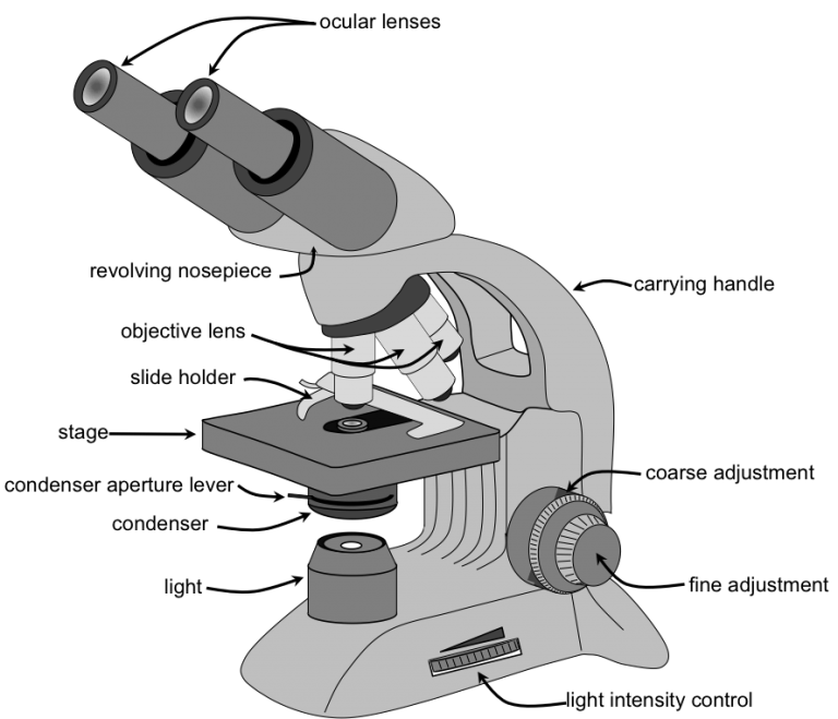
- Eyepiece – The eyepiece contains the ocular lens. It’s found at the top of the microscope. Its standard magnification is 10X with an optional eyepiece having magnifications from 5X – 30X. The eyepiece contributes to the magnification of the object on stage.
- Eyepiece tube – This is the eyepiece holder. It carries the eyepiece just above the objective lens. In some microscopes such as the binoculars, the eyepiece tube is flexible and can be rotated for maximum visualization, for variance in distance. For monocular microscopes, they are none flexible.
- Objective lenses – There are generally 3 or 4 objective lenses in a microscope. They almost always consist of 4x, 10x, 40x and 100x powers. The objective lens gathers light from the specimen, magnifies the image of the specimen, and projects the magnified image into the body tube. Since no single objective lens can fulfill all the needs of someone using the microscope, several objective lenses of varying magnification and numerical aperture are mounted on the rotating nosepiece. The nosepiece must click into place for the objective lens to be in proper alignment. You will notice that the lower power lenses (4X and 10X) are shorter than the high power lenses (40X and 100X), meaning that the clearance between the objective lens and the stage is much smaller when the high power lenses are clicked into place. You must be very careful when using the high power lenses so you do not jam them into the slide.
- Revolving Nose piece – also known as the revolving turret. Several objective lenses of varying magnification and numerical aperture are mounted on the revolving nosepiece. The main function of revolving nose piece is to hold and bring the objective lens in position.
- The Adjustment knobs – There are two types of adjustment knobs i.e fine adjustment knobs and the coarse adjustment knobs.
- The coarse adjustment knob- This knob is located on the arm of the microscope moves the stage up and down to bring the specimen into focus. The gearing mechanism of the adjustment produces a large vertical movement of the stage with only a partial revolution of the knob. Because of this, the coarse adjustment should only be used with low power (4X and 10X objectives) and never with the high power lenses (40X and 100X).
- The fine adjustment knob- This knob is inside the coarse adjustment knob and is used to bring the specimen into sharp focus under low power and is used for all focusing when using high power lenses.
- Stage – The flat horizontal platform upon which the slide is placed is called the stage. The slide is held in place by spring loaded clips and moved around the stage by turning the geared knobs on the stage. The stage has two perpendicular scales that can be used to record the position of an object on a slide. This is useful if you want to quickly relocate an object. The stage also has a central slit that allows light passing from the illuminator and condenser to penetrate the specimen.
- Microscopic illuminator – This is the microscopes light source, located at the base. It is used instead of a mirror. It captures light from an external voltage bulb of about 100v. Older microscopes used mirrors to reflect light from an external source up through the bottom of the stage; however, most microscopes now use a low-voltage bulb.
- Condenser Lens – The purpose of the condenser lens is to focus/concentrate the light onto the specimen. They are found under the stage next to the diaphragm of the microscope. Condenser lenses are most useful at the highest powers (400x and above). Microscopes with in-stage condenser lenses render a sharper image than those with no lens (at 400X). The higher the magnification of the condenser, the more the image clarity.
- Diaphragm or Iris – Many microscopes have a rotating disk under the stage. This diaphragm has different sized holes and is used to vary the intensity and size of the cone of light that is projected upward into the slide. There is no set rule regarding which setting to use for a particular power. Rather, the setting is a function of the transparency of the specimen, the degree of contrast you desire and the particular objective lens in use.
- Aperture – This is a central slit or hole on the microscope stage, through which the transmitted light from the source reaches the stage.
- Condenser focus knob – This control is used to precisely adjust the vertical height of the condenser.
- Abbe Condenser – A lens that is specially designed to mount under the stage and which typically moves in a vertical direction. An adjustable iris controls the diameter of the beam of light entering the lens system. Both by changing the size of this iris and by moving the lens toward or away from the stage, the diameter and focal point of the cone of light that goes through the specimen can be controlled. Abbe condensers are useful at magnifications above 400X where the condenser lens has a numerical aperture equal to or greater than the N.A. of the objective lens being used.
- The rack stop – This is an adjustment that determines how close the objective lens can get to the slide. Setting the rack stop is useful in preventing the slide from coming too far up and hitting the objective lens. Normally, this adjustment is set at the factory, and changing the rack stop is only necessary if your slides are exceptionally thin and you are unable to focus the specimen at higher powers.
- Field Diaphragm Control – The base of the microscope contains the field diaphragm. This controls the size of the illuminated field. The field diaphragm control is located around the lens located in the base.
- Hinge Screw-This screw fixes the arm to the base and allow for the tilting of the arm.
- Stage Clips– They hold the slide firmly onto the stage.
- On/OFF Switch– This switch on the base of the microscope turns the illuminator off and on.
How To Use A Light Microscope
To View the Object
- Turn the low power objective lens until it clicks into position
- Looking through the eye piece, ensure that enough light is passing through by adjusting the mirror
- This is indicated by a bright circular area known as the field of view
- Place the slide containing the specimen on stage and clip it into position
- Make sure that the specimen is in the centre of the field of view
- Using the coarse adjustment knob, bring the low power objective lens to the lowest point
- Turn the knob gently until the specimen comes into focus
- If finer details are required, use the fine adjustment knob
- When using high power objective always move the fine adjustment knob upwards
Handling And Care Of A Microscope
- Great care should be taken when handling it.
- Keep it away from the edge of the bench when using it.
- Always hold it with both hands when moving it in the laboratory.
- Clean the lenses with special lens cleaning paper.
- Make sure that the low power objective clicks in position in line with eye piece lens before and after use.
- Store the microscope in a dust-proof place free of moisture.