Gram Positive Bacteria

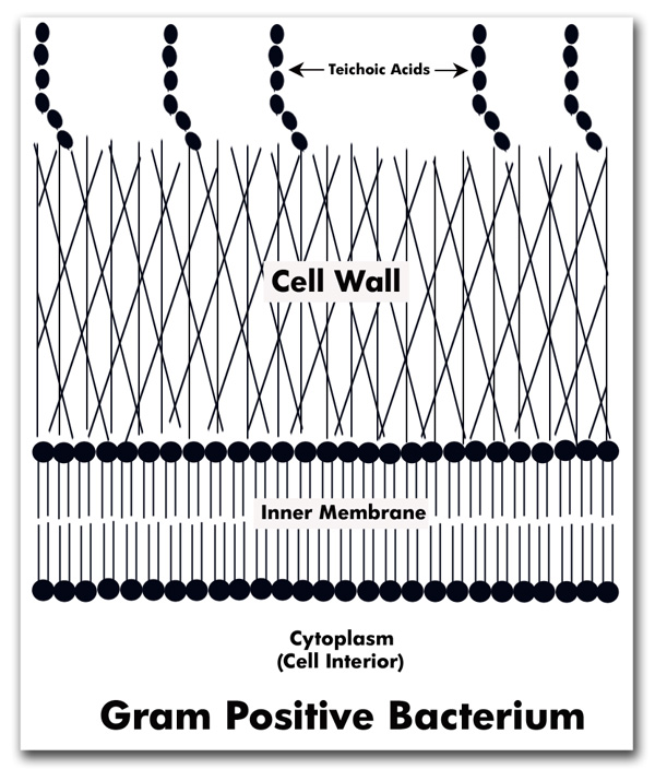
Gram-positive bacteria are bacteria with thick cell walls. In a Gram stain test, these organisms yield a positive result. The test, which involves a chemical dye, stains the bacterium’s cell wall purple.
Gram-positive bacteria take up the crystal violet stain used in the test, and then appear to be purple-coloured when seen through an optical microscope. This is because the thick peptidoglycan layer in the bacterial cell wall retains the stain after it is washed away from the rest of the sample, in the decolorization stage of the test.
The most common gram negative bacteria include:
- Staphylococcus epidermidis
- Streptococcus pneumoniae
- Streptococcus pyogenes
- Streptococcus agalactiae
- Bacillus anthracis
- Corynebacterium diphtheria
- Listeria monocytogenes

Gram Negative Bacteria

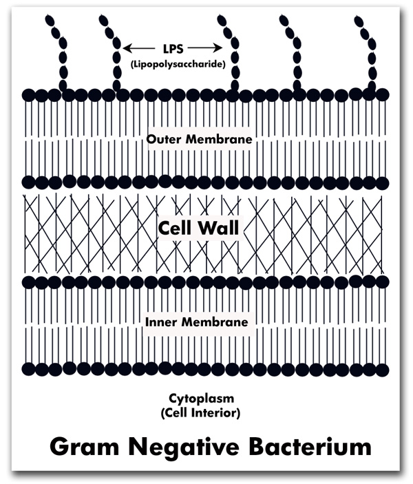
Gram-negative bacteria are bacteria that do not retain the crystal violet stain used in the gram-staining method of bacterial differentiation.They are characterized by their cell envelopes, which are composed of a thin peptidoglycan cell wall sandwiched between an inner cytoplasmic cell membrane and a bacterial outer membrane.
Gram-negative bacteria can cause many serious infections, such as pneumonia, peritonitis (inflammation of the membrane that lines the abdominal cavity), urinary tract infections, bloodstream infections, wound or surgical site infections, and meningitis
The most common Gram Negative Bacteria Include:
- Escherichia,
- Klebsiella,
- Pseudomonas,
- Serratia
- Enterobacter.

Cell Wall Anatomy


Key Differences
| Thickness of cell wall | Cell wall of gram positive bacteria is 20 to 80 nanometer thick | Cell wall of gram negative bacteria is 8 to 10 nanometer thin |
| Cell Wall Peptidoglycan layer | Smooth single layered cell wall Peptidoglycan layer is thick up to 90% and are multilayered | Wavy double-layered cell wall Peptidoglycan layer is thin and are often single layered |
| Teichoic acid | Teichoic acid is present within the cell wall | Teichoic acid is absent |
| Periplasmic space | There is no presence of periplasmic space | Periplasmic space is present between the region of cytoplasmic and outer membrane |
| Outer membrane | Outer membrane is absent but are surrounded by thick layers of peptidoglycan | Outer membrane is mostly present with a unique component, lipopolysaccharide (LPS) with proteins and phospholipids |
| Rigidity | Gram positive cell wall are more rigid due to cross linkage of peptidoglycan chains | Gram negative cell wall are less rigid |
| Elasticity | Cell wall is less elastic | Cell wall is more elastic |
| Color observed on Gram’s staining | Purple or blue colored is observed when examined under the microscope after Gram’s staining which is due to the retaining of crystal violet dye by the cell wall of gram positive bacteria. | Pink or red colored observed when examined under the microscope after Gram’s staining which accepts the counterstain safranin by the cell wall of gram negative bacteria after decolourization step. |
| Morphology of bacteria | Cocci or spore forming rods Gram positive cocci: Streptococcus pneumonia, Staphylococcus aureus, Enterococcus Gram positive rods: Corynebacterium diphtheria, Listeria monocytogenes, Bacillus anthracis, Erysipelothrix rhusiopathiae | Cocci or non-spore forming rods Gram negative cocci: Neisseria gonorrhoeae, Neisseria meningitides, Moraxella catarrhalis, Haemophilus influenza Gram negative rods: Escherichia coli, Yersinia pestis, Salmonella Typhi/Paratyphi, Salmonella, Shigella, Proteus |
| Porins | Not present | Present in outer membrane that helps in the diffusion of small hydrophilic molecules |
| Mycolic acid | Present in some bacteria like Mycobacterium tuberculosis that makes up to 60% of the cell wall | Absence of mycolic acid |
| Mesosome | Mesosomes are more prominent | Mesosomes are less prominent |
| Lipoprotein content | Acid fast bacteria have lipids connected to peptidoglycan layer therefore, lipoprotein content is low | Due to presence of outer layer, lipoprotein content is high |
| Toxins produced | Exotoxins | Either exotoxins or endotoxins |
| Flagellar structure | Two rings in basal body, one in the peptidoglycan layer and one in the plasma membrane | Four rings in basal body: L ring in the lipopolysaccharide layerP ring in the peptidoglycan layerM ring embedded in the plasma membraneS ring directly connected to the plasma membrane |
| Lipid content | Very low and are imbedded in the cytoplasmic membrane | Up to 20 to 30% in the outer membrane |
| Endospore formation | Formation of endospore during unfavourable conditions (eg: Bacillus cereus, Bacillus anthracis, Bacillus thuringiensis Clostridium botulinum, Clostridium tetani) | Usually endospores are not formed |
| Cell wall disruption by lysozyme | Gram positive bacteria are more susceptible towards the lysozyme action because of their cell wall content up to 90% peptidoglycan layer | Gram negative bacteria are more resistant towards lysozyme due to its low content of peptidoglycan layer |
| Resistance to physical disruption | Gram positive bacteria are more resistant to physical disruption | Gram negative bacteria are less resistant |
| Resistance to antibiotics | Most gram positive bacteria are sensitive to antibiotics | They are more resistant to antibiotics because of the presence of large impermeable cell wall |
| Antibiotics used to treat bacterial infections | Vancomycin, teicoplanin, Quinupristin, Oxazolidinones, Daptomycin, Telavancin, Ceftaroline | Cephalosporins, fluoroquiolones, aminoglycosides, imipenem, broad spectrum penicillins with or without β-lactamase inhibitors |
| Resistance to drying | Highly resistant towards drying due to their thicker cell wall | Low resistant towards drying |
| Resistance to disinfectants | More susceptible to cleaning agents due to lack of an outer membrane | Less susceptible to cleaning agents due to the reduced permeability of the double membrane |
| Nutritional Requirements | Nutritional requirement is relatively complex | Nutritional requirement is relatively simple |
| Inhibition by basic dyes like salts, chlorides | Highly inhibited by basic dyes | Low inhibited by basic dyes |
| Susceptibility to anionic detergents like Sodium n-dodecyl benzene sulphonate and Sodium Lauryl sulphate | Highly susceptible towards anionic detergents | Less susceptible towards anionic detergents |
| Murein content | Murein content in cell wall is high about 70 to 80% | Murein content in cell wall is low about 10 to 20% |
| Variety of amino acids in cell wall | Few variety of amino acids are found in the cell wall of gram positive bacteria | Several variety of amino acids are found in the cell wall of gram negative bacteria |
| RNA:DNA ratio | In gram positive bacteria, the ratio of RNA:DNA is 8:1 | In gram negative bacteria, the ratio of RNA:DNA is 1-3:1 which is almost 1 |
| Pathogens | Only six gram positive genera are pathogenic to humans | Most gram negative bacteria are pathogenic to humans |
| Diseases | Pharyngitis, cellulitis, scarlet fever, necrotizing fasciitis, glomerulonephritis, rheumatic fever. | Brucellosis, Cholera. Plague, Typhoid fever, urinary tract infections (UTI), bloodstream infections, meningitis. |